3D Echo – Your Frequently Asked Questions Answered
Last year we launched 3D Echo, the new module for HeartWorks.
In this blog, Consultant Cardiac Anaesthetists, Dr. Sue Wright MBBS, FRCA, Dr. Andrew Smith MBBS, FRCA and Dr. Bruce Martin MBBS, FRCA answer your Frequently Asked Questions (FAQ) on one of the latest additions to the Intelligent Ultrasound family:
What is live 3D cardiac ultrasound?
Live 3D cardiac ultrasound entails the generation of a moving 3D image of the heart.
What is 3D echocardiography used for in a clinical setting?
In a clinical setting 3D scanning allows us to understand complex cardiac anatomy. The ability to do this in real time allows us to use 3D echo to guide complex cardiac procedures that may be either cardiac surgery or structural interventional procedures in the cardiac catheter labs.
What are the benefits of learning/practising on a simulator vs in-clinic?
Learning how to acquire 3D datasets is both difficult and time consuming. Although different ultrasound vendors use different terminology and workflows to acquire 3D datasets, there are many common principles in the process. The ability to gain an understanding of these principles as they apply to image acquisition and manipulation techniques in a way that is not dependent on clinical time gives students an ideal opportunity to learn the skills required for 3D echocardiography.
How do you switch to 3D scanning in HeartWorks?
3D imaging is accessed in HeartWorks by selecting the 3D button from the imaging mode icons on the simulator console. Entering the 3D imaging mode opens context-appropriate options for dataset acquisition and manipulation.
How do you perform the 3D scan in HeartWorks?
Upon entering 3D mode in HeartWorks the user will be given the choice of full volume or zoom data acquisition. The simulator will take the student through the steps of data acquisition, offering choices with respect to breath hold and multibeat acquisition as in real-life scanning. Acquired datasets can be manipulated and cropped, and MPR planes can be generated from them.
How can you use the 3D scan?
The simulated datasets will allow detailed anatomical inspection of cardiac anatomy and function, as in real life. Dataset acquisition appropriate to real time procedure guidance can also be achieved.
What are the main benefits of HeartWorks 3D Echo to a clinician?
- The ability to learn common principles of 3D dataset acquisition
- The ability to learn cropping
- The ability to understand factors that affect image resolution and clarity
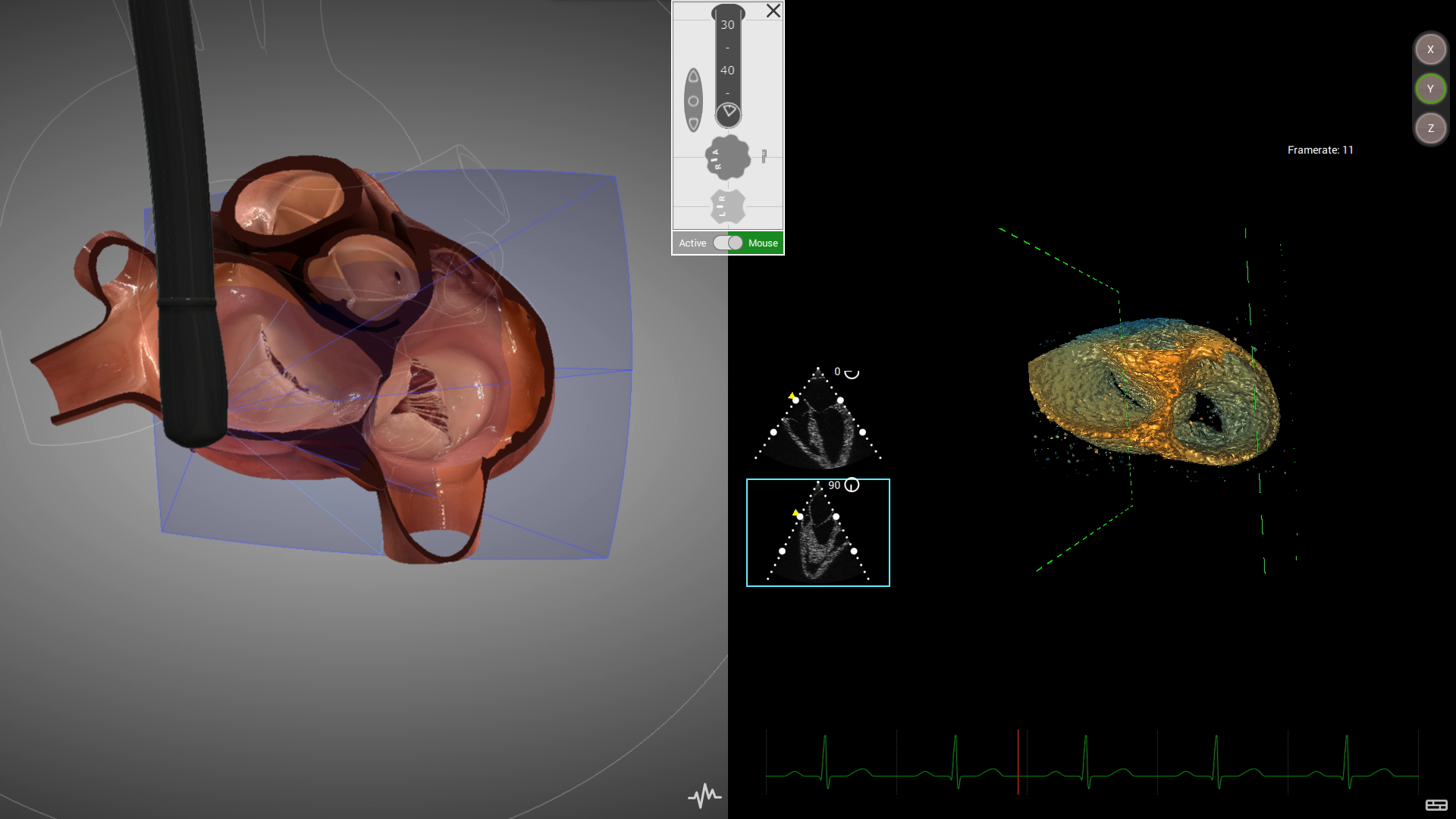
- The ability to see in the 3D heart model how the ultrasound frustum is positioned relative to cardiac structures
For more information on HeartWorks and 3D Echo, click here.